
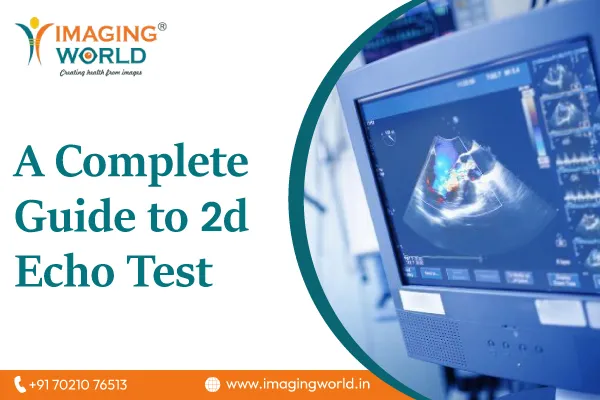
2D echo of the heart also known as 2D Echocardiography is a diagnostic method wherein ultrasound waves are used to create a picture of the heart in the human body. The picture is a cross-sectional slice representation of the heart with its beats, picture of chambers, valves, and all the primary blood vessels. Doppler is a key element of the 2d echo test in Ahmedabad which is used to assess the blood flow in the heart. This is the reason this test is also known as Doppler ultrasound of the heart.
Why is the 2d echo of heart used?
Echocardiography or 2d Echo is used by physicians to extract a range of important information about a patient’s heart which includes:
1. The volume and thickness of the walls along with the size of the chambers
2. Whether the pumping function of a heart is normal or affected to a mild or severe degree
3. The normality of the valve function including structure, movement and thickness of the valves
4. Volume status of the heart. A common outcome of poor heart function can be low blood pressure
5. Test of the fluid in the pericardium, clinically known as Pericardial Effusion
6. Any congenital heart disease
7. Blood clots and tumors
8. Abnormal elevation of pressure in the lungs
Procedure of 2d Echo test:
To perform the 2d echo test in Ahmedabad, the patient is asked to change to a robe which is open at the front. A colorless gel is applied to the chest area of the patient. The patients are instructed to lay on his left side to allow full access to the technician to move a transducer across the various parts of the chest. The patient is asked to breathe as directed, slow/ fast, as per the requirement of the scan. The images are viewed on a monitor and are recorded in a CD or Paper.
How long does it take to get results from a Doppler test?
The procedure does not take more than 20-25 minutes. But, in certain cases, as per the condition of the heart, it may take longer than the mentioned time limit.
How does a Doppler ultrasound work?
In a Doppler ultrasound test, ultrasound waves are used to create a picture of the organs. It is a non-invasive method which is used to estimate the flow of blood in various organs including the heart, arms and legs. Color Doppler in pregnancy is often used to identify the flow of blood in the fetus. The high-frequency ultrasound waves are reflected off of the various organ’s blood flow, which helps in the testing.
Color Doppler is an effective test that allows physicians to get a clear view of the flow of blood in various organs. Thanks to the evolving technology, the color Doppler test cost has also become much cheaper than it was a few years back. The test is known to be absolutely safe which no reported side-effects. However, the results are quoted as highly superior as the traditional ultrasound tests.