
Ultrasound imaging – also referred to as sonography is a medical procedure often carried out in hospitals using high frequency sound waves to map images within the body.
Technology and Medical Research
Medical scientists and physicians are constantly conducting research and testing new procedures to help prevent, diagnose, and cure diseases as well as developing new drugs and medicines that can lessen symptoms or treat ailments.
Ultrasound is a non invasive procedure and unlike X-Ray which uses potentially harmful ionizing radiation, ultrasound is a virtually risk free procedure which is why it is used routinely to scan images of unborn babies.
Colour Doppler Ahmedabad
The other additional benefit of ultrasound is that it captures images in real time and can show movement within the body, making it ideal to monitor blood flow within the body.
General ultrasound images are produced in 2d but advances in the technology have meant that images can now be processed in 3d and in some very special instances 4d (movement is recorded)
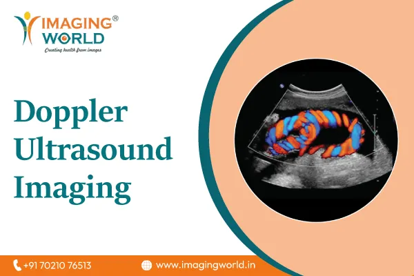
Doppler ultrasound is a specialized technique which evaluates the flow of blood around the body as it moves through the body’s major veins and arteries.
Color Doppler test uses Doppler measurements to create a color pattern which describes the flow of blood through the blood vessel it is measuring.
Power Doppler is used to give detailed information of blood flow within deeper blood vessel – located within organs in the body.
Spectral Doppler displays Doppler measurements graphically instead of using color patterns.
Common Ultrasound Procedures Ultrasound scanning has a wide spectrum of medical some very common uses for it are to diagnose swelling and infection within the body.
It is also extremely well suited to examining internal organs including:
Heart, liver, gallbladder , spleen, pancreas, kidneys, bladder uterus, ovaries, eyes,thyroid
Another use for Ultrasound scanning is for guiding a needle thought the body during biopsies (to extract a sample of body cells).
Common Doppler procedures
Some routine Doppler ultrasound procedures
Evaluate blockages within blood vessels (blood clots) Evaluate the narrowing of blood vessels caused by plaque
This information can be used to determine whether a patient would be suitable for a procedure like angioplasty.
What to do if you are having an ultrasound examination
Loose fitting clothing should be worn if you are about to have an ultrasound examination, your physician will ask you to remove your clothes and any jeweler and you will be wear a gown when the procedure is taking place.
In some more specialized ultrasound examinations your doctor may tell you not to eat or drink for a period before your examination. But generally this is not the case for routine examinations.
What does ultrasound equipment look like? An ultrasound scanner consists of a computer and video display, the actual transducer that scans the body looks a bit like a microphone which is attached to the computer console by a cord.
The transducer sends out sound waves and records the returning echo which is then used to create the image. The basic principles are the same as sonar used in submarines and ships.