
 MRI • CT • Sonography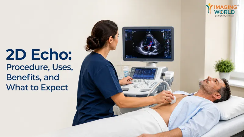
Key Takeaways
- A 2D Echo is a non-invasive ultrasound test that provides real-time images of the heart's structure and function.
- Understanding the 2D Echo procedure helps alleviate anxiety and makes the visit smoother for patients.
- Doctors recommend a 2D Echo in Ahmedabad for symptoms like unexplained shortness of breath and chest pain.
- The test offers crucial insights into heart chambers, valves, and pumping function without any ionising radiation.
- Patients should prepare by bringing previous medical records and following their doctor's advice before the 2D Echo.
Imaging World is a leading diagnostic imaging centre providing 2D Echo services for heart assessment. Your heart works every second of the day, yet changes in its structure or pumping function may require detailed medical evaluation. When a doctor needs real-time information about heart movement, chambers, valves, and blood flow patterns, echocardiography can be an important diagnostic investigation.
Ultrasound waves are used in a 2D echocardiography to produce moving pictures of the heart. Ionizing radiation is not used in this non-invasive test. For patients advised to undergo a 2D Echo in Ahmedabad, knowing the procedure, uses, benefits, and what happens during the examination can make the visit more comfortable and easier to understand.
What Is a 2D Echo?
A 2D Echo, also known as two-dimensional echocardiography, is an ultrasound-based imaging test used to examine the heart. It creates real-time images that allow healthcare professionals to assess the structure and movement of different parts of the heart.
The test may provide information about:
- Heart chambers
- Heart valves
- Heart wall movement
- Pumping function
- Heart size
- Certain blood flow patterns when Doppler assessment is included
- Fluid around the heart
Unlike an X-ray or CT examination, echocardiography works with sound waves. Your doctor may recommend the test based on symptoms, clinical findings, medical history, or the need to monitor a known heart condition.
Why Is a 2D Echo Recommended?
Doctors use echocardiography to gather visual information about heart structure and function. The test is not limited to a single heart problem. Its role depends on the patient's clinical condition and the questions the treating doctor needs to evaluate.
A doctor may advise a 2D Echo in Ahmedabad when a patient has symptoms or findings such as:
- Unexplained shortness of breath
- Chest-related symptoms requiring cardiac evaluation
- Swelling in the legs
- Abnormal heart sounds or a heart murmur
- Suspected heart valve problems
- Concerns about heart pumping function
- Follow-up of certain known cardiac conditions
These symptoms do not automatically confirm heart disease. The treating doctor considers examination findings and other investigations before making a diagnosis.
How Does the 2D Echo Procedure Work?
The procedure is generally simple from the patient's point of view. During a standard transthoracic echocardiogram, you may be asked to remove clothing from the upper body as required and wear a suitable covering.
Small electrodes may be placed to monitor the heart rhythm during the examination. You will usually lie on an examination couch, often partly turned onto your left side.
A trained professional applies ultrasound gel to the chest and moves a handheld device called a transducer over selected areas. The transducer sends and receives sound waves, allowing the ultrasound system to create images of the beating heart.
During the 2D Echo, you may occasionally be asked to change position or briefly control your breathing. This can help obtain the required imaging views. The examination itself is non-invasive, although mild pressure from the transducer may sometimes be felt.
What Can a 2D Echo Show?
One major benefit of echocardiography is its ability to show the heart in motion. Instead of providing only a still structural view, it allows assessment of how different heart structures move during the cardiac cycle.
The examination may help assess:
Heart Chamber Size
The images can help evaluate the dimensions of the heart chambers and identify enlargement when present.
Pumping Function
Echocardiography can provide information about how effectively the heart is contracting and pumping blood.
Heart Valves
The test allows assessment of valve structure and movement. Doppler techniques may also provide information about blood flow across valves.
Heart Wall Movement
The movement of different heart wall segments can be examined as part of the cardiac assessment.
Pericardial Fluid
The test may identify fluid collection around the heart when present.Patients searching for a 2D Echo in Bopal should remember that the clinical interpretation of these findings must be performed by qualified medical professionals and considered alongside the patient's overall health information.
Key Benefits of a 2D Echo
Echocardiography is widely used in cardiac evaluation because it provides useful real-time information without ionising radiation.
Some practical benefits include:
- Non-invasive examination: A standard transthoracic echo does not involve surgical instruments.
- Real-time heart imaging: Doctors can assess heart structures while the heart is beating.
- No ionising radiation: Ultrasound sound waves are used to create images.
- Structural assessment: Heart chambers, valves, and wall movement can be examined.
- Functional information: The test can support evaluation of heart pumping and blood flow characteristics.
- Useful for follow-up: Doctors may use repeat echocardiography to monitor selected cardiac conditions.
At Imaging World, we understand that cardiac investigations can make some patients feel concerned. Clear guidance about the examination can help patients know what to expect during their visit.
Do You Need to Prepare for a 2D Echo?
A standard transthoracic echocardiogram usually requires little special preparation. However, patients should adhere to the advice given by their physician or diagnostic facility.
Before visiting for a 2D Echo in Ahmedabad, it is useful to carry:
- Your doctor's prescription or referral
- Previous cardiac reports, if available
- Previous echo reports for comparison, when relevant
- A list of current medicines if requested
- Other related medical records advised by your doctor
Do not stop prescribed medicines unless your treating doctor specifically tells you to do so.Preparation can differ for other forms of echocardiography, so always confirm the instructions for the exact test recommended to you.
What Should You Expect After the Test?
After a standard 2D echocardiogram, the ultrasound gel is cleaned from the chest and most patients can generally return to their usual activities unless their doctor has given different instructions.
The captured images are reviewed and interpreted by the relevant medical professional. Your treating doctor will consider the echo findings together with your symptoms, medical history, examination, and other investigations.
Imaging World focuses on providing a professional diagnostic experience where patients receive appropriate guidance during the imaging process. We believe that good communication is particularly important when someone visits for a heart-related investigation.
Choosing a Centre for Your 2D Echo
When looking for a 2D Echo in Bopal, patients should consider the quality of diagnostic services rather than treating the examination as a routine formality. Proper imaging technique and professional interpretation play an important role in cardiac assessment.
Consider factors such as qualified professionals, suitable ultrasound technology, patient guidance, a clean examination environment, and an organised reporting process.
A 2D Echo in Bopal may be recommended as part of an initial cardiac assessment or follow-up plan. Always share relevant previous reports and clinical information when requested, as this can provide useful context for medical evaluation.
Why Choose Us
Imaging World, focuses on providing professional diagnostic imaging services with attention to patient guidance and quality examination processes. We understand that patients visiting for cardiac imaging may have questions about the test and its purpose, so our approach centres on clear communication and a comfortable diagnostic experience. Our team follows appropriate imaging procedures while focusing on obtaining clinically useful images for medical assessment. Imaging World is dedicated to providing patients with responsible diagnostic services and a professional approach to cardiac imaging, regardless of whether you have been recommended to have an echo for initial evaluation or follow-up.
Conclusion
A 2D echocardiogram is a valuable non-invasive imaging test that provides real-time information about heart structure, valves, chambers, wall movement, and pumping function. Doctors may recommend it for symptoms, clinical findings, known cardiac conditions, or follow-up assessment. The procedure is generally straightforward and uses ultrasound rather than ionising radiation. Understanding what happens before, during, and after the examination can help patients feel better prepared. Imaging World provides professional cardiac imaging support with a focus on patient guidance and quality diagnostic processes. Always discuss your echo findings with your treating doctor for proper clinical interpretation and further medical advice.
Need a professional 2D Echo for heart assessment? Book your appointment with Imaging World today. Call +91 70210 76513 or Book Your Appointment for diagnostic imaging assistance.